Redium Diagnostic Center

""Redium Never compromised with quality any Test""

Redium Diagnostic Center

""Redium Never compromised with quality any Test""

Redium Diagnostic Center

""Redium Never compromised with quality any Test""

Redium Diagnostic Center

""Redium Never compromised with quality any Test""

Redium Diagnostic Center

""Redium Never compromised with quality any Test""

গ্যাস্ট্রোএন্টেরোলজিস্ট ও মেডিসিন বিশেষজ্ঞ (কাপাসিয়া শাখা)


ডাঃ রনজিত কুমার পাল
এমবিবিএস, বিসিএস (স্বাস্থ্য), এমডি (গ্যাস্ট্রোএন্টেরোলজি)
এফসিপিএস-এফ (মেডিসিন), ডিএলপি (ডায়াবেটোলজি, বারডেম)
নিউরোলজি, হৃদরোগ, বাত ব্যথা, বক্ষব্যাধি কিডনী রোগে উচ্চতর প্রশিক্ষণ প্রাপ্ত ইন্টারভেশনাল গ্যাস্ট্রোএন্টেরোলজি-হেপাটোলজিষ্ট।
সহকারী অধ্যাপক বিভাগীয় প্রধান (গ্যাস্ট্রোলিভার মেডিসিন বিভাগ)
শহীদ তাজউদ্দিন আহমদ মেডিকেল কলেজ হাসপাতাল, গাজীপুর।

রোগী দেখার সময় :
প্রতি শুক্রবার সকাল ১০.৩০টা থেকে দুপুর .০০টা  মঙ্গবার .৩০ টা থেকে বিকাল .০০টা পর্যন্ত।


ডাঃ মাহবুবুর রহমান
এমবিবিএস, আই এইচ এমএড (ইরান) এমসিপিএস, ডিএসএম (গ্যাস্ট্রো)
এফ এম এস (অষ্টিয়া), এম জি এইচ (ইউকে) এম এফ সি (আমেরিকা)
এম ডি (গ্যাস্ট্রো), এফ সি পি এস (মেডিসিন)
ফেলো অব রয়েল সোসাইটি অব হেলথ (লন্ডন)
আজীবন মেম্বার আমেরিকান মেডিকেল সোসাইটি

রোগী দেখার সময়:
প্রতি রবিবার সকাল ১০.৩০টা থেকে বিকাল .০০টা পর্যন্ত


ডাঃ মোঃ সাইয়েদুল আশিক (শাওন)
এমবিবিএস (মমেক), বিসিএস (স্বাস্থ্য)
এমডি (হেপাটোলজি)-বিএসএমএমহউ
সিসিডি (বারডেম)
ইন্টারভেনশনাল হেপাটো গ্যাস্ট্রোএন্টেরোলজিস্ট
শহীদ তাজউদ্দিন আহমদ মেডিকেল কলেজ হাসপাতাল, গাজীপুর।

রোগী দেখার সময়:
প্রতি সোমবার দুপুর .৩০টা থেকে বিকাল .০০টা পর্যন্ত।


Imaging Tests

Different Types of Imaging Tests

Radium Diagnostic Center has several types of imaging tests, each of which has its own specific purpose and advantages. Here are some of the most common types:

X-ray :

           This is the most common type of imaging test, which uses radiation to create images of the inside of the body. X-rays are often used to diagnose bone fractures and other skeletal injuries, as well as to detect abnormalities in the chest and abdomen.

Computed Tomography (CT) scan :

           A CT scan uses X-rays and computer technology to create detailed images of the body. This type of test is often used to diagnose conditions such as cancer, heart disease, and brain disorders.

Magnetic Resonance Imaging (MRI):

           MRI uses a strong magnetic field and radio waves to create detailed images of the inside of the body. This type of test is often used to diagnose conditions such as tumors, internal bleeding, and joint injuries.


Ultrasound:

          This type of imaging uses sound waves to create images of the inside of the body. It is often used to diagnose conditions such as pregnancy, liver disease, and heart disease.

 Positron Emission Tomography (PET) scan:

         A PET scan uses a radioactive substance to create images of the body. It is often used to diagnose cancer, heart disease, and brain disorders.


Mammography:

        Mammography uses X-rays to create images of the breasts. It is often used to screen for breast cancer.

 Fluoroscopy:

           This type of imaging uses X-rays to create real-time images of the inside of the body. It is often used during medical procedures such as surgeries and catheterizations.

These are just a few examples of the many types of imaging tests available. Each test has its own advantages and limitations, and the choice of which test to use will  depend on the specific condition being diagnosed and other factors. 

Click Here for Contact

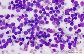
Acute Lymphoblastic Leukaemia


What is Acute lymphoblastic leukaemia

Acute lymphoblastic leukemia (ALL), also known as acute lymphocytic leukemia, is a type of cancer that affects the blood and bone marrow. It is a rapidly progressing form of leukemia that affects the white blood cells known as lymphocytes, which are responsible for fighting infections in the body.

In ALL, immature lymphocytes called lymphoblasts grow and divide uncontrollably, crowding out healthy blood cells and interfering with their function. This can lead to a range of symptoms, including fatigue, weakness, frequent infections, easy bruising or bleeding, bone pain, and swollen lymph nodes.

ALL is the most common type of leukemia in children, but it can occur in people of any age. Treatment for ALL typically involves a combination of chemotherapy, radiation therapy, and other targeted therapies, with the goal of killing cancer cells and restoring healthy blood cell production. The outlook for people with ALL has improved significantly in recent years, with high rates of remission and long-term survival, particularly in younger patients.


Warning signs of acute leukaemia :

The warning signs of acute leukemia can vary depending on the type of leukemia and the individual. Some of the common warning signs of acute leukemia include:

1.             Fatigue and weakness: Feeling very tired or weak, even after getting enough rest, is a common symptom of leukemia.

2.             Frequent infections: People with leukemia may have a weakened immune system, which can make them more susceptible to infections.

3.             Easy bruising or bleeding: Leukemia can affect the production of blood cells, which can lead to easy bruising or bleeding.

4.             Bone pain or tenderness: Leukemia can cause pain or tenderness in the bones, particularly in the limbs and joints.

5.             Swollen lymph nodes: Lymph nodes can become enlarged and feel tender or painful.

6.             Unexplained weight loss: People with leukemia may lose weight without trying to.

7.             Shortness of breath: Leukemia can affect the production of red blood cells, which can lead to shortness of breath.

8.             Headaches: Leukemia can cause headaches or other neurological symptoms, particularly in cases where the cancer has spread to the central nervous system.

If you are experiencing any of these symptoms, it is important to speak with your doctor as soon as possible to determine the cause and receive appropriate treatment.

What happens in acute leukaemia :

In acute leukemia, abnormal white blood cells, also called blasts, grow and divide rapidly in the bone marrow, interfering with the production of normal blood cells. These abnormal cells do not function properly, and they accumulate in the bloodstream, bone marrow, and other organs, eventually leading to symptoms and complications.

Acute leukemia can develop quickly over a matter of weeks or months, and it can affect both children and adults. There are two main types of acute leukemia: acute lymphoblastic leukemia (ALL) and acute myeloid leukemia (AML).

In ALL, the abnormal white blood cells are immature lymphocytes, which are a type of white blood cell that normally helps fight infections. In AML, the abnormal white blood cells are immature myeloid cells, which are the cells that give rise to red blood cells, white blood cells, and platelets.

The rapid growth and accumulation of these abnormal cells can lead to a variety of symptoms, including fatigue, weakness, frequent infections, easy bruising or bleeding, bone pain, swollen lymph nodes, and more. Treatment for acute leukemia typically involves chemotherapy, radiation therapy, and other targeted therapies to kill the cancer cells and restore normal blood cell production.


Acute cholecystitis


About Acute cholecystitis

Acute cholecystitis is a condition where there is inflammation of the gallbladder, typically caused by the blockage of the cystic duct by gallstones. The gallbladder is a small, pear-shaped organ located just below the liver that stores bile, a digestive fluid produced by the liver. When the gallbladder becomes inflamed, it can cause severe abdominal pain, fever, and nausea.

The symptoms of acute cholecystitis usually develop suddenly and may include:

·         Severe abdominal pain, especially in the upper right part of the abdomen

·         Nausea and vomiting

·         Fever

·         Jaundice (yellowing of the skin and eyes)

·         Loss of appetite

·         Tenderness in the abdomen, particularly over the gallbladder

If you suspect you have acute cholecystitis, it is important to seek medical attention promptly, as the condition can lead to serious complications such as a ruptured gallbladder or infection. Treatment may involve antibiotics, pain relief, and in severe cases, surgical removal of the gallbladder.

What causes acute cholecystitis?

Acute cholecystitis is most commonly caused by gallstones, which are small, hard deposits that form in the gallbladder. When a gallstone becomes lodged in the cystic duct, it can block the flow of bile, causing inflammation and swelling of the gallbladder. This can lead to the symptoms of acute cholecystitis, including severe abdominal pain, fever, and nausea.

Other less common causes of acute cholecystitis include:

·  Tumors or growths in the gallbladder

·   Infection in the gallbladder

·  Trauma or injury to the gallbladder

·  Biliary sludge, which is a mixture of cholesterol and other substances that can form in the gallbladder

Risk factors for developing acute cholecystitis include being female, over the age of 40, obese, having a family history of gallstones or gallbladder disease, and having certain medical conditions such as diabetes or cirrhosis of the liver.


Diagnosing cholecystitis

The diagnosis of cholecystitis typically involves a combination of medical history, physical examination, and imaging tests. The following are some of the diagnostic tests commonly used to diagnose cholecystitis:

1. Medical history: The doctor will ask you about your symptoms, medical history, and any medications you are taking.

2.  Physical examination: The doctor will perform a physical examination to check for signs of inflammation, tenderness, and pain in the upper right part of your abdomen.

3.   Blood tests: Blood tests can be done to check for signs of infection, inflammation, and liver function.

4.     Ultrasound: An ultrasound is a non-invasive imaging test that uses sound waves to create images of the gallbladder. It is often the first imaging test performed in the diagnosis of cholecystitis.

5.    CT scan: A CT scan is a more detailed imaging test that can provide a more detailed picture of the gallbladder and surrounding structures.

6.   HIDA scan: A HIDA scan is a nuclear medicine test that can evaluate the function of the gallbladder and detect any blockages or inflammation.

If you are experiencing symptoms of cholecystitis, it is important to seek medical attention promptly to determine the cause of your symptoms and receive appropriate treatment.

Treating acute cholecystitis

The treatment of acute cholecystitis depends on the severity of the condition and may involve medical management or surgical intervention. Here are some common treatments:

1.   Nonsurgical Treatment: Mild cases of cholecystitis may be treated with medications to control pain and inflammation, and antibiotics to treat any underlying infection. Nonsurgical treatment can also be used to stabilize patients who are not fit for surgery or those with milder symptoms.

2.   Surgical Treatment: Surgical intervention is often recommended for severe or recurrent cases of acute cholecystitis. The most common surgery for cholecystitis is laparoscopic cholecystectomy, a minimally invasive surgical procedure in which the gallbladder is removed using small incisions in the abdomen. Open cholecystectomy, a more invasive surgical procedure, may be recommended in cases where laparoscopic surgery is not feasible or in cases of severe inflammation.

3.  Diet modification: In addition to medical or surgical treatment, dietary changes may also be recommended to manage symptoms. A low-fat diet is often recommended to reduce the workload of the gallbladder.

It is important to seek medical attention promptly if you suspect you have acute cholecystitis, as the condition can lead to serious complications such as a ruptured gallbladder or infection. The treatment plan will depend on the severity of your symptoms and the underlying cause of your condition, and will be determined by your healthcare provider.

 Preventing acute cholecystitis

While it is not always possible to prevent acute cholecystitis, there are some measures you can take to reduce your risk of developing the condition:

1.  Maintain a healthy weight: Being overweight or obese can increase your risk of developing gallstones, which are a common cause of cholecystitis. Maintaining a healthy weight through regular exercise and a healthy diet can help reduce your risk.

2.  Eat a healthy diet: Eating a diet that is low in fat and high in fiber can help reduce your risk of developing gallstones. Foods that are high in cholesterol or saturated fats should be avoided or consumed in moderation.

3.  Stay hydrated: Drinking plenty of water can help prevent the formation of gallstones, as well as reduce the risk of inflammation and infection in the gallbladder.

4. Manage underlying medical conditions: Certain medical conditions such as diabetes or liver disease can increase your risk of developing gallstones and cholecystitis. Managing these conditions through regular medical care and treatment can help reduce your risk.

5.  Avoid rapid weight loss: Rapid weight loss, such as that caused by crash dieting or bariatric surgery, can increase the risk of developing gallstones and cholecystitis. Gradual weight loss through a healthy diet and exercise is recommended.

It is important to talk to your healthcare provider about any concerns you may have about your risk of developing cholecystitis and to follow their recommendations for preventive measures.